3D Imaging - Techniques Improve Clinical Examination For Orthodontists
3D imaging was created in the early 1990s and has since established a valuable role in dentistry, particularly in orthodontics.
Author:Suleman ShahReviewer:Han JuJul 18, 20239.1K Shares338.6K Views

3D imagingwas created in the early 1990s and has since established a valuable role in dentistry, particularly in orthodontics.
Orthodontic and dentofacial orthopedic diagnosis and treatment planning have traditionally depended heavily on technical and mechanical aids such as imaging, jaw monitoring, and functional evaluations.
These approaches aim to perfectly duplicate or explain anatomic and physiological facts and portray three-dimensional (3D) anatomy.
One of the essential tools for orthodontists to examine and record the size and shape of craniofacial features is imaging.
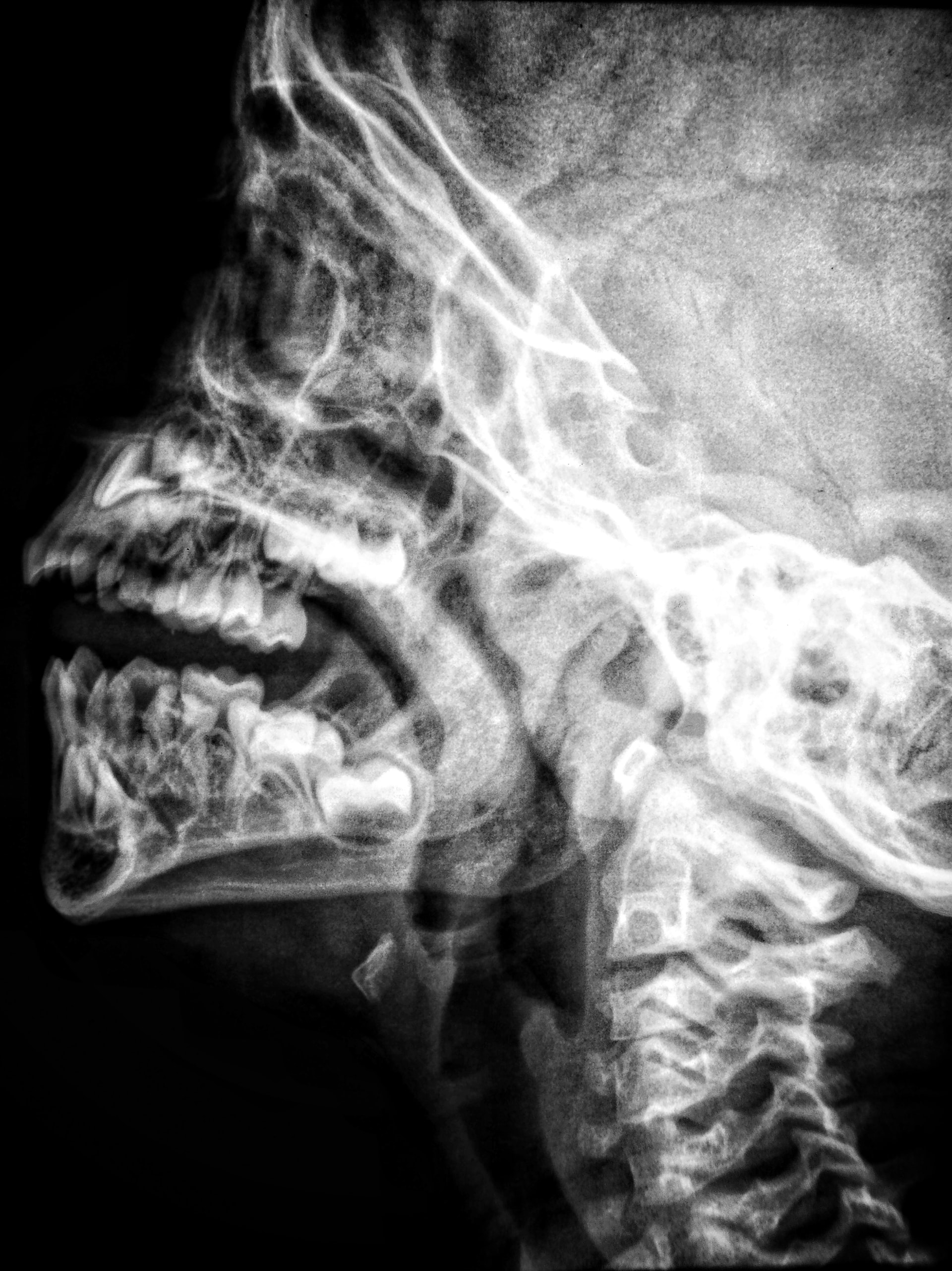
Orthodontists regularly employ 2-dimensional (2D) static imaging methods to capture craniofacial anatomy, yet 2D imaging cannot collect and locate structural depth.
3D imaging was created in the early 1990s and has since established a valuable role in dentistry, particularly in orthodontics and orofacial surgical applications.
In 3D diagnostic imaging, anatomical data is collected using specific technical equipment, analyzed by a computer, and then shown on a 2D monitor to create a sense of depth.
How Does 3D Imaging Work?
Stereography is used in 3D imaging, as we can see from a familiar source: the human visual system.
Humans perceive with two eyes that are somewhat apart.
This enables individuals to experience depth in addition to the horizontal and vertical information represented by a normal 2D television screen, for example.
Because the eyes are separated, each observes the world differently.
Covering one eye quickly, the other reveals minor but noticeable variations in angle each time.
Human vision has dimensionality because the brain combines diverse pictures into a whole, known as parallax.
Every 3D photo employs two lenses, each of which takes an image that is slightly offset from the other.
As a consequence, 3D photographs have double the information as 2D photos.
The photos are altered to ensure complete data accuracy when displaying.
Each eye processes its own set of pictures. Hence the eye cannot process both sets of images simultaneously.
In the brain, the left-hand and right-hand pictures merge to form a sensation of depth.
3D Imaging Techniques
Computed Tomography
Computed tomography (CT) imaging, also known as Computerized Axial Tomography (CAT) imaging, creates cross-sections of the body using specialized X-ray equipment.
The CT scanner produces a narrow, fan-shaped beam of X-rays that scans a part of the patient's body.
Although specific tests need a contrast substance to help certain portions of the body show more clearly in the picture, the technique is painless.
In advanced fan-beam CT, 64 and 128 sections may be achieved simultaneously.
CT scans yield a large amount of data, including 3D information on soft and hard tissues.
Furthermore, when compared to other soft-tissue imaging modalities, CT data is inadequate.
Cone Beam Computerized Tomography (CBCT)
Craniofacial CBCT devices are intended to address some of the shortcomings of traditional CT scanning technologies.
They emit 15 times less radiation than traditional CT scanners.
Realignment of 2-dimensional pictures in coronal, sagittal, oblique, and different inclination planes is possible with CBCT.
The radiation exposure of CBCT is equivalent to the dose of 12 panoramic radiographs.
In personal computers, CBCT may display and organize 3D data.
There are several sophisticated software available for implant implantation and orthodontic measures.
CBCT not only has a limited capability for exhibiting soft tissues, but it also has a space for investigating hard tissueof the head and face.
Previous research found that when the CBCT was used, the incidence of oral abnormalities increased compared to previous studies.
The Nasopharyngeal Airway Analysis
As a result, either surgical removal of the adenoids/ tonsils or obstructive sleep apnea therapydue to narrow airways can be applied if necessary.
In both studies, airways were found to be unchanged after orthodontic treatment.
Cleft Lip/Palate Patients (CL/P Patients)
Because Cleft Lip/ Palate (CL/P) is so common in the community, it is not unexpected that studies on CBCT imaging in orofacial abnormalities have focused on these individuals.
To ensure adequate alveolar bone in CL/P patients, the appropriate quantity of graft material may be created using CBCT volumetric studies.
Based on the discrepancies in soft tissue measures from CBCT pictures across the three groups, synchronous rhinoplasty is recommended to improve the nasal and labial look.
Temporomandibular Joint (TMJ) Morphology
Using 3-dimensional software, images of cranial structures collected at various periods may be placed on pre-defined spots.
The CBCT picture may reveal changes in the look of the face caused by tooth movement, orthognathic surgery, or other craniofacial therapy.
Furthermore, 3D Fotoscan machines may be used to create models of CBCT pictures.
Micro-Computed Tomography (MCT)
MCT is similar to CT, except the reconstructed cross-sections are limited to a considerably smaller region.
Conventional CT scans capture 0.012 mm thin cross-sections, whereas MCT can be acquired with nano-sized sections.
Mineralized tissues are analyzed using MCT, a non-invasive and non-destructive approach.
MCT's future resides in its ability to collect input across a much smaller volume than the whole body, thus minimizing radiation exposure.
3D Laser Scanning
Laser scanning provides 3D pictures for treatment planning or analyzing the results of orthodontic and specifically orthognathic therapy as a less intrusive means of capturing the face.
Furthermore, 3D laser scanners may generate digital models.
Structured Light Technique
Structured light scanning allows for the easy and non-ionizing 3-dimensional contour of the face.
The first 3D hand-held intra-oral scanner, Ora-Scanner, is based on structured light methods.
Techalertpaisan and Kuroda created a 3D representation of a face form that could be adjusted, moved, or rotated softly in all directions using two LCD projectors, a camera, and a computer.
This technique needs at least 2 seconds to acquire a picture, which may be too long to prevent baby and toddler movements.
Sterophotogrametry
Stereophotogrammetry is the process of photographing a three-dimensional object from two separate coplanar planes.
In the face display, this strategy has shown to be quite successful.
Thalmann-Degan performed the first clinical use of stereophotogrammetry in 1944, recording the changes on the face due to orthodontic therapy.
Stereophotogrammetry for 3D photos using two separate coplanar planes: Tissue reflections, hair and brow intervention, changing posture between views, and motions during imaging reduce the likelihood of acquiring the most accurate face pictures.
3D Facial Morphometry (3DFM)
The system comprises two infrared cameras, hardware for marker detection, and software for 3D reconstruction of landmark coordinates.
The placement of landmarks on the face is time-consuming and labor-intensive.
This technique cannot generate models with the genuine soft-tissue look of a facial expression.
Magnetic Resonance Imaging (MRI)
Radio wavesare directed to the required place for analysis in a magnetic field.
The hydrogen nucleus is used to provide a resonance signal for MRI.
The energy generated by hydrogen atoms in radio-stimulated cells is transformed into numbers.
They are turned into images after being processed on a computer.
People Also Ask
What Is 3D Imaging Techniques?
3D imaging is a technology that provides the illusion of depth inside a picture by converting 2D data into a 3-dimensional format.
3D imaging has become a handy feature in quality controlling procedures for industrial applications.
How Does 3D Medical Imaging Work?
Ultrasonographers utilize a probe to evaluate a patient's anatomy using 3D ultrasound.
They take 3D image sweeps and crucial pictures and transfer them to a 3D workstation.
Before the pictures are sent to the radiologist, a 3D ultrasound technician evaluates them and generates additional 3D views.
What Is 3D Imaging In Dentistry?
Dentists use a special scanner called a Cone Beam Computed Tomography (CBCT) machine to take photos of your teeth and jaw.
This equipment helps dentists look at your teeth by producing a 3D picture.
Conclusion
The need has pushed the development of 3D imaging for high speed, high density, compact size, and multipurpose devices.
New imaging methods need costly software and a significant amount of time.
Robotic devices that are quicker and more versatile seem to be the future of 3D imaging.

Suleman Shah
Author
Suleman Shah is a researcher and freelance writer. As a researcher, he has worked with MNS University of Agriculture, Multan (Pakistan) and Texas A & M University (USA). He regularly writes science articles and blogs for science news website immersse.com and open access publishers OA Publishing London and Scientific Times. He loves to keep himself updated on scientific developments and convert these developments into everyday language to update the readers about the developments in the scientific era. His primary research focus is Plant sciences, and he contributed to this field by publishing his research in scientific journals and presenting his work at many Conferences.
Shah graduated from the University of Agriculture Faisalabad (Pakistan) and started his professional carrier with Jaffer Agro Services and later with the Agriculture Department of the Government of Pakistan. His research interest compelled and attracted him to proceed with his carrier in Plant sciences research. So, he started his Ph.D. in Soil Science at MNS University of Agriculture Multan (Pakistan). Later, he started working as a visiting scholar with Texas A&M University (USA).
Shah’s experience with big Open Excess publishers like Springers, Frontiers, MDPI, etc., testified to his belief in Open Access as a barrier-removing mechanism between researchers and the readers of their research. Shah believes that Open Access is revolutionizing the publication process and benefitting research in all fields.

Han Ju
Reviewer
Hello! I'm Han Ju, the heart behind World Wide Journals. My life is a unique tapestry woven from the threads of news, spirituality, and science, enriched by melodies from my guitar. Raised amidst tales of the ancient and the arcane, I developed a keen eye for the stories that truly matter. Through my work, I seek to bridge the seen with the unseen, marrying the rigor of science with the depth of spirituality.
Each article at World Wide Journals is a piece of this ongoing quest, blending analysis with personal reflection. Whether exploring quantum frontiers or strumming chords under the stars, my aim is to inspire and provoke thought, inviting you into a world where every discovery is a note in the grand symphony of existence.
Welcome aboard this journey of insight and exploration, where curiosity leads and music guides.
Latest Articles
Popular Articles
